- sales@hot-mining.com +86 28 8331 1885 +86 15196677188
-

-

-


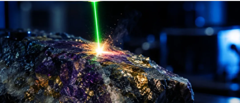
Laser-Induced Breakdown Spectroscopy (LIBS) has become a widely used technique for elemental and mineralogical imaging of rocks and minerals. However, not all LIBS imaging instruments are designed in the same way. The underlying system architecture can have a direct impact on data accuracy.
Many LIBS imaging systems currently available on the market use what I would describe as a "semi-sequential spectrometer architecture." These systems combine Czerny-Turner spectrometers with ICCD cameras, with each unit responsible for a relatively narrow spectral window. At high resolution, this window is typically 10 nm; at lower resolutions, it may extend to 20 or 40 nm.
The economic cost of this design creates a practical constraint.
A single Czerny-Turner/ICCD unit costs approximately USD 135,000. Covering a meaningful portion of the periodic table with this approach would require two or three such units, putting the total cost beyond the reach of many laboratories and service providers.
As a result, many instruments can only observe part of the emission spectrum at any given time.
To cover a wider range of elements, these systems must scan the same sample surface multiple times, with each scan configured for a different spectral window.
The issue lies in the fact that LIBS is a semi-invasive technique.
Each laser pulse ablates approximately 1–5 µm of material from the sample surface.
For single-point elemental analysis, this is generally not a problem. For imaging and quantitative automated mineralogy, however, it can become a significant concern.
The structures of rocks and minerals are three-dimensional and are often heterogeneous at the micrometre scale.
When a semi-sequential instrument first images Layer 1, then rescans the sample to capture Layer 2, and finally scans it again for Layer 3, it is not actually imaging the same surface each time. Instead, it is imaging progressively deeper regions of the sample, which have already been affected by previous laser scans.
The more spectral windows that need to be covered, the more scans are required. With each successive scan, the sample surface moves further away from its original state.
As a result, the elemental and mineralogical distribution maps generated from Spectral Windows 1, 2, and 3 do not actually represent the same physical layer. They describe information from different depths within the sample, yet are combined as if they came from one continuous and consistent image.
This introduces systematic bias into the spectral data and can affect any subsequent quantitative analysis or mineralogical interpretation. Moreover, as the number of target elements increases, this bias may become more pronounced rather than being eliminated.
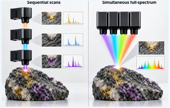
Figure: Comparison of sequential LIBS imaging (limited spectral window of 10 nm) and simultaneous LIBS imaging (e.g., 215–950 nm).
An alternative system architecture uses multiple CMOS-based Czerny-Turner spectrometers operating in coordination to capture the complete atomic emission spectrum simultaneously and without gaps, in a single laser pulse, across an approximate wavelength range of 215–950 nm.
For rock and mineral imaging, this approach offers two direct advantages.
Direct Advantage 1: No Resampling Artifacts
Because each pixel's complete spectrum is acquired in a single measurement, every image layer corresponds to the same physical depth within the sample.
There is no depth mismatch between spectral channels caused by cumulative ablation, nor are there errors associated with positional changes, such as sample movement or backlash-related positioning errors in the translation stage.
Direct Advantage 2: Richer Information from a Single Acquisition
Gap-free, full-spectrum acquisition captures not only atomic emission lines but also continuum emission and molecular bands.
These signals may be missed by systems that divide the spectrum into narrow windows, or may be captured inconsistently across multiple scans.
If you are evaluating a LIBS imaging platform for geological or mineralogical research, one question deserves particular attention:
Can the instrument acquire the full spectral range in a single scan, or does it need multiple repeated scans to cover different elements?
If multiple scans are required, it is also important to understand how the supplier addresses the resulting challenge: data acquired during successive scans may no longer represent the same physical depth.
As the laser continues to ablate the sample, the surface changes continuously. This can introduce depth-related discrepancies between different spectral windows.
Therefore, the accuracy of elemental and mineralogical imaging is not determined solely by spectral resolution.
The more fundamental question is whether the data ultimately combined and analyzed genuinely originate from the same location and the same physical layer within the sample.